


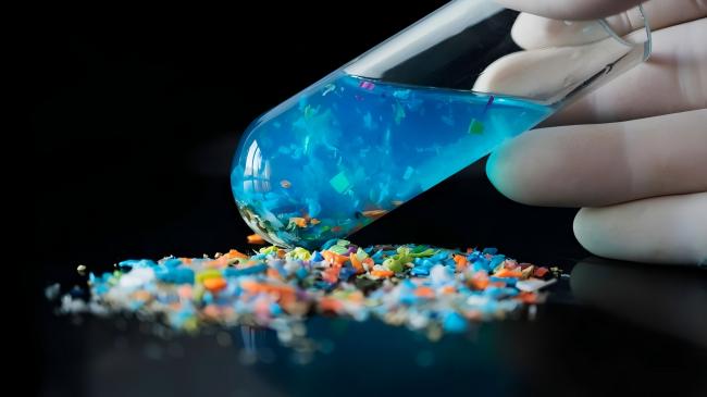
Microplastic Detection
Identify and quantify microplastic particles in water with advanced analytical techniques combining microscopy, spectroscopy, and automated imaging systems. Our facilities in Korea, Singapore, and China deliver comprehensive microplastic characterization including particle counting, size distribution, polymer identification, and morphological classification. From drinking water to wastewater, surface water to marine environments, our detection services address the growing concern of plastic pollution in aquatic ecosystems and drinking water supplies.
Understanding Microplastic Contamination
The Global Microplastic Crisis
Why microplastics are emerging as critical water contaminants:
- What are Microplastics? - Plastic particles <5 mm in size (WHO/GESAMP definition) | Nanoplastics: <1 µm (1,000 nm) | Primary microplastics: manufactured small (microbeads in cosmetics, industrial abrasives, pre-production pellets) | Secondary microplastics: degradation of larger plastic items (bottles, bags, textiles, tires) | Composed of various polymers: polyethylene (PE), polypropylene (PP), polystyrene (PS), polyethylene terephthalate (PET), polyvinyl chloride (PVC), polyamide (PA/nylon)
- Sources in Water Systems - Wastewater treatment plant effluent (incomplete removal, 90-99% retained but millions of particles still discharged daily) | Stormwater runoff (tire wear particles, degraded litter, microbeads) | Industrial discharges (textile manufacturing, plastic production, recycling facilities) | Atmospheric deposition (airborne microplastics settle into water bodies) | Breakdown of larger plastic debris in rivers, lakes, oceans | Agricultural runoff (plastic mulch films, irrigation systems) | Synthetic textile washing (fibers shed during laundry cycles, 700,000+ fibers per wash)
- Environmental Persistence & Transport - Extremely persistent - plastics degrade over decades to centuries | Fragmentation continues indefinitely (microplastics → nanoplastics) | Buoyant plastics (PE, PP) travel long distances in surface waters | Denser plastics (PET, PVC) settle in sediments | Biofilm formation alters density and transport behavior | Vertical mixing transports microplastics throughout water column | Long-range transport via ocean currents, rivers (microplastics detected in Arctic, deep ocean trenches)
- Human Health Concerns - Ingestion via drinking water (WHO estimates: 0-4 particles/L in treated drinking water, higher in bottled water) | Potential for translocation across gut barrier (particles <10 µm) | Chemical toxicity from plastic additives (phthalates, bisphenols, flame retardants) leaching in digestive system | Physical effects from particle accumulation (inflammation, oxidative stress) | Vector for pathogen transport (bacteria, viruses colonize microplastic surfaces) | Adsorption of environmental contaminants (POPs, heavy metals, pharmaceuticals) - microplastics as Trojan horses | Long-term health effects largely unknown - emerging research area
- Ecological Impacts - Ingestion by aquatic organisms (zooplankton to whales) - mistaken for food | Bioaccumulation and biomagnification through food webs | Physical harm (gut blockage, reduced feeding, false satiation) | Toxic effects from leached additives on reproduction, development | Disruption of nutrient cycling in aquatic ecosystems | Habitat alteration (microplastics in sediments change substrate properties) | Species-specific impacts (filter feeders particularly vulnerable)
- Regulatory Landscape - No established drinking water limits yet (WHO states no health risk at current levels but data limited) | EU developing regulatory framework for microplastics in environment | California AB 1066 (2018): requires monitoring of microplastics in drinking water | Research and monitoring requirements expanding globally | GESAMP (UN) harmonizing measurement methodologies | Increasing focus on source control (microplastic bans in cosmetics, improved wastewater treatment)
Analytical Challenges for Microplastic Detection
Why Microplastic Analysis is Complex
Unique challenges requiring specialized equipment and expertise:
- Sample Preparation Complexity - Large sample volumes needed (10-100L for drinking water, 1-10L for wastewater) | Concentration via filtration (multiple filter sizes: 5 mm, 1 mm, 300 µm, 100 µm, 50 µm, 20 µm, 5 µm) | Removal of natural organic matter (algae, detritus, biofilms obscure microplastics) - enzymatic or chemical digestion required | Density separation (saturated salt solutions: NaCl, NaI, ZnCl₂) to float plastics away from sediment | Filtration artifacts (filter material contamination, particle losses during transfers) | Extreme care to avoid contamination from laboratory plastics, clothing, air
- Detection & Identification Difficulties - Visual sorting inadequate (misidentification of non-plastic particles, observer bias, time-consuming) | Chemical confirmation required for every suspected particle | Size range spans 5 orders of magnitude (5 mm to 1 µm or smaller) | Irregular shapes (fibers, fragments, films, foams, beads, pellets) complicate automated detection | Transparent and colorless particles difficult to visualize | Degraded/weathered plastics have altered surface properties | No single technique detects full size range and all polymer types
- Polymer Identification - Requires spectroscopic analysis: FTIR (Fourier-Transform Infrared) or Raman spectroscopy | FTIR: particles >50-100 µm (diffraction limit), provides polymer fingerprint, library matching (10,000+ polymer spectra) | Raman: particles >1 µm, laser-based, excellent for small particles but fluorescence interference common | Micro-FTIR: imaging mode for spatial mapping of polymer distribution | Raman microscopy: high spatial resolution but slow for large particle numbers | Pyrolysis-GC/MS: destructive technique, identifies polymer type and additives but no particle morphology
- Quantification Challenges - Report as particles per liter (p/L) or mass per liter (mg/L or µg/L) | Particle counting: labor-intensive, requires imaging each particle | Mass determination: difficult for small particles, aggregates complicate weighing | Size distribution: requires image analysis of hundreds to thousands of particles | Variability between replicates (spatial heterogeneity in water samples) | Detection limit depends on sample volume, smallest filter size, and analytical technique
- Contamination Control - Airborne microplastics ubiquitous (synthetic clothing fibers, dust) | All laboratory consumables potential sources (plastic bottles, pipette tips, tubing, gloves) | Strict protocols required: cotton lab coats, glass/metal equipment only, HEPA-filtered clean benches, positive air pressure labs | Procedural blanks mandatory (>10% of samples) - air blanks, filtration blanks, digestion blanks | Contamination typically 0-5 particles per blank (subtracted from sample counts)
FTIR Spectroscopy (Chemical Identification)
Micro-FTIR (Transmission Mode) - Particles >100 µm analyzed individually | Infrared beam passes through particle on KBr window or diamond compression cell | Generates IR spectrum (fingerprint of polymer functional groups) | Library matching against 10,000+ polymer spectra (>80% match threshold) | Identifies: PE, PP, PS, PET, PVC, PA, PC, and additives | Analysis time: 1-3 min per particle (manual positioning)
FTIR Microscopy (Attenuated Total Reflectance, ATR) - Particles 50-5,000 µm | Diamond ATR crystal in contact with particle | No sample preparation, non-destructive | Better for irregular/thick particles than transmission mode | Spatial resolution: ~10-20 µm
FTIR Imaging (Focal Plane Array, FPA) - Simultaneously images 64×64 pixel array (~5 µm pixel size) | Entire filter scanned automatically (hyperspectral imaging) | Each pixel generates IR spectrum → polymer map | Identifies and counts 1,000+ particles per filter | Analysis time: 4-24 hours per filter (size dependent) | Gold standard for comprehensive microplastic analysis | Commercial systems: Agilent 8700 LDIR, PerkinElmer Spotlight, Bruker Lumos
Raman Spectroscopy
Raman Microscopy (Confocal) - Particles >1 µm (excellent for small microplastics) | Laser excitation (532 nm, 785 nm) generates Raman scatter | Highly specific polymer identification | Spatial resolution: ~0.5-1 µm (better than FTIR) | Limitations: Fluorescence interference (pigments, degraded plastics), sample heating with high laser power, slow (2-5 min per particle)
Raman Imaging - Automated stage scans entire filter | Generates Raman spectrum at each pixel | Polymer maps identify distribution and composition | Excellent for colored/pigmented microplastics (FTIR struggles with dark particles) | Analysis time: 8-48 hours per filter (slower than FTIR imaging)
Surface-Enhanced Raman (SERS, Experimental) - Gold/silver nanoparticles enhance Raman signal 10⁶-10⁸× | Detects nanoplastics (<100 nm) | Research technique, not yet routine
Pyrolysis-GC/MS
Thermal Desorption GC/MS - Bulk sample (filter) heated to 300-700°C in pyrolyzer | Plastics decompose to characteristic monomers/oligomers | GC separates pyrolysis products, MS identifies them | Quantifies total polymer mass (µg to mg) | Identifies polymer types and plastic additives simultaneously | Advantages: High sensitivity, analyzes entire sample, detects additives (phthalates, BPA, flame retardants) | Disadvantages: Destructive (no particle morphology), no individual particle data | Commercial systems: Frontier Lab Multi-Shot Pyrolyzer, Gerstel TDS-GC/MS
Emerging Techniques
Hyperspectral Imaging (Vis-NIR) - Visible and near-infrared spectroscopy (400-2,500 nm) | Rapid imaging (faster than FTIR/Raman) | Identifies major polymer types but less specific than FTIR/Raman | Good for large particles (>500 µm)
Atomic Force Microscopy (AFM) - Nanoscale imaging (<100 nm particles) | Surface topography and mechanical properties | Identifies nanoplastics but slow and expensive | Research applications
Electron Microscopy + EDX - SEM: Morphology at nm resolution | EDX: Elemental composition (identifies chlorine in PVC, etc.) | Identifies weathered/degraded microplastics by surface features | Requires conductive coating (limits spectroscopy follow-up)
Typical Microplastic Concentrations in Water
Drinking Water, Surface Water, and Wastewater
Concentration ranges from global monitoring studies:
| Water Type | Sample Matrix | Concentration Range (particles/L) |
Dominant Polymer Types | Common Particle Shapes |
|---|---|---|---|---|
| Drinking Water (Treated) | Tap water from municipal systems | 0-4 (WHO estimate) 0.1-10 (published studies) |
PE (30-40%), PP (20-30%), PET (15-25%), PS (5-15%) | Fibers (60-70%), fragments (20-30%), films (5-10%) |
| Bottled Water | Commercial bottled water (plastic bottles) | 10-6,000 (avg: 100-300) |
PET (40-60%, from bottle), PP (20-30%, from cap), PE (10-20%) | Fragments (50-60%), fibers (30-40%), films (5-10%) |
| Groundwater | Wells, aquifers | 0.1-5 (limited data) |
PE (30-40%), PP (20-30%), PET (15-25%) | Fibers (50-60%), fragments (30-40%) |
| Rivers (Freshwater) | Surface water, subsurface | 0.5-50 (highly variable by location) |
PE (35-45%), PP (25-35%), PS (10-20%), PET (5-15%) | Fragments (40-50%), fibers (35-45%), foams (5-10%) |
| Lakes & Reservoirs | Surface water (drinking water sources) | 1-40 (lower in remote lakes) |
PE (30-40%), PP (20-30%), PET (15-25%), PS (10-20%) | Fibers (50-60%), fragments (30-40%), films (5-10%) |
| Coastal/Estuarine Waters | Surface water near coastlines | 5-200 (higher near urban areas) |
PE (40-50%), PP (25-35%), PS (10-20%), PET (5-10%) | Fragments (45-55%), fibers (30-40%), foams (10-15%) |
| Open Ocean (Surface) | Pelagic waters, gyres | 0.1-100 (gyres: 10-1,000+) |
PE (50-70%, buoyant), PP (20-30%), PS (5-10%) | Fragments (60-70%), foams (15-25%), films (10-15%) |
| Wastewater (Influent) | Raw sewage entering treatment plant | 100-10,000 (avg: 500-2,000) |
PE (30-40%), PP (20-30%), PET (20-30%, textile fibers), PA (5-15%) | Fibers (70-85%, laundry), fragments (10-20%), beads (2-5%) |
| Wastewater (Effluent) | Treated wastewater discharge | 1-100 (90-99% removal) |
PE (30-40%), PP (20-30%), PET (15-25%), PA (10-15%) | Fibers (65-75%), fragments (20-30%), films (5-10%) |
| Stormwater Runoff | Urban stormwater, combined sewer overflows | 50-5,000 (highly variable, event-dependent) |
PE (30-40%), PP (20-30%), PET (15-25%), tire rubber (10-20%) | Fragments (50-60%), fibers (25-35%), tire particles (10-15%) |
Important Notes: Concentrations vary widely depending on analytical method (filter size, identification technique). Smaller filters detect more particles. Studies using <10 µm filters report 10-100× higher counts than >100 µm filters. Polymer percentages approximate - vary by region and water type. Tire wear particles (styrene-butadiene rubber) increasingly recognized as major microplastic source but often excluded from surveys (not true plastics).
Microplastic Size Distribution
Particle abundance by size class (general pattern across water types):
| Size Class | Size Range | Relative Abundance | Detection Method | Characteristics |
|---|---|---|---|---|
| Large Microplastics | 1-5 mm | 5-15% | Visual sorting + FTIR/Raman | Easily visible, fragmented plastic debris, pellets, large fibers |
| Medium Microplastics | 300 µm - 1 mm | 15-25% | Stereomicroscope + micro-FTIR | Visible with magnification, textile fibers, paint chips, foam fragments |
| Small Microplastics | 50-300 µm | 25-40% | Optical microscope + FTIR imaging | Require microscopy, microfibers, tire dust, degraded fragments |
| Very Small Microplastics | 10-50 µm | 30-50% | High-mag microscopy + Raman imaging | Near limit of optical microscopy, secondary degradation products |
| Nanoplastics (Large) | 1-10 µm | Unknown (under-sampled) |
Raman microscopy, pyrolysis-GC/MS | Difficult to distinguish from inorganic particles, require advanced techniques |
| Nanoplastics (Small) | <1 µm (1,000 nm) | Unknown (rarely measured) |
AFM, SERS, TEM, pyrolysis-GC/MS | Likely most abundant numerically, research frontier, health concern (cellular uptake) |
Key Observation: Abundance increases exponentially with decreasing size. For every particle 1-5 mm, there may be 10-100 particles 50-300 µm, and thousands to millions of particles <10 µm. Most studies underestimate total microplastic load due to limited detection of small particles.
Analytical Workflows
Standard Microplastic Analysis Protocol
Comprehensive workflow for water samples:
Step 1: Sample Collection & Filtration
Sample Volume: 10-100L (drinking water), 1-10L (wastewater), 0.5-5L (surface water with high MP load)
Collection: Stainless steel or glass containers, no plastic contact
Filtration: Cascade filtration through 5 mm → 1 mm → 300 µm → 50 µm stainless steel sieves, final filtration through GFF (1.2 µm) for small particles
Storage: Filters stored in aluminum foil or glass petri dishes at -20°C until analysis
Time: 1-4 hours depending on volume and turbidity
Step 2: Organic Matter Removal
Enzyme Digestion: Filters incubated with Proteinase K (10 mg/mL) at 50°C for 24 hours in glass beakers on orbital shaker
Oxidation (if needed): Fenton's reagent (30% H₂O₂ + 0.05 M Fe²⁺) at 50°C for 24 hours for heavily contaminated samples
Rinse: Filters rinsed with ultrapure water, back-filtration to collect digested particles
QC: Digestion blank processed simultaneously
Time: 24-48 hours
Step 3: Density Separation (Optional)
Salt Solution: NaCl (ρ=1.2 g/cm³) or ZnCl₂ (ρ=1.6 g/cm³) depending on target plastics
Procedure: Resuspend filter contents in salt solution in separatory funnel, settle 12-24 hours, collect floating layer
Repeat: 2-3× to maximize plastic recovery (>95%)
Filtration: Re-filter floating fraction onto clean GFF or stainless steel mesh
Time: 2-3 days (mostly hands-off)
Step 4: Microscopy & Particle Identification
Visual Inspection: Stereomicroscope screening at 10-50× magnification, suspected microplastics marked for analysis
Imaging: High-resolution images of each particle (size, shape, color documentation)
Optional Staining: Nile Red fluorescence for rapid plastic vs. non-plastic screening
Particle Counting: Manual count or automated image analysis
Time: 2-8 hours per filter depending on particle density
Step 5: Polymer Identification
FTIR Analysis: Micro-FTIR for particles >100 µm (1-3 min each) or FTIR imaging for entire filter (4-24 hours)
Raman Analysis: Raman microscopy for particles 1-100 µm (2-5 min each) or Raman imaging (8-48 hours)
Library Matching: Compare spectra to polymer databases, >80% match threshold for positive ID
Result: Polymer type for each particle (PE, PP, PS, PET, PVC, PA, etc.)
Time: Hours to days depending on method and particle number
Step 6: Data Analysis & Reporting
Blank Correction: Subtract average blank count from sample counts
Normalization: Calculate particles per liter (p/L) based on sample volume
Size Distribution: Histogram of particle sizes
Polymer Composition: Pie chart or bar graph of polymer types
Morphology: Classify as fibers, fragments, films, foams, beads
Report Generation: Comprehensive data package with images, spectra, statistics
Rapid Screening Protocol (Pyrolysis-GC/MS)
Bulk analysis for total microplastic quantification:
Pyrolysis-GC/MS Workflow
Sample Preparation: Entire filter (or portion) placed in pyrolyzer sample cup, no individual particle isolation needed
Pyrolysis: Rapid heating to 600-700°C in inert atmosphere (helium), plastics decompose to monomers (e.g., styrene from PS, terephthalic acid from PET)
GC/MS Analysis: Pyrolysis products separated by GC, identified and quantified by MS
Quantification: Total mass of each polymer type (µg to mg) based on calibration with polymer standards
Advantages: Rapid (30 min per sample), high sensitivity (ng detection limits), identifies additives simultaneously
Limitations: No particle count, no size distribution, no morphology, destructive
Best For: Screening large numbers of samples, total microplastic burden assessment, wastewater monitoring
Thermal Desorption Profile
Polymers Identified: PE, PP, PS, PET, PVC, PA, PC, ABS, PMMA, and others based on pyrolysis markers
Additives Detected: Phthalates (plasticizers), bisphenols (BPA, BPS), flame retardants (PBDE, HBCD), UV stabilizers, antioxidants
Quantification Range: 0.1 µg to 10 mg polymer per filter
Detection Limits: 0.05-0.5 µg polymer mass (equivalent to ~10-1,000 particles depending on size)
Applications: High-throughput screening, method comparison (validates imaging results), regulatory monitoring when particle-level data not required
Service Packages
Basic Microplastic Screening
Analysis Scope: Particles >100 µm, visual identification + FTIR confirmation for suspected plastics
Included: Filtration (5 mm, 1 mm, 300 µm, 100 µm cascade), organic matter digestion (enzymatic), microscopy screening, micro-FTIR on 50-100 particles/sample
Deliverables: Total particle count (p/L), polymer types (% of identified particles), size distribution, morphology classification, images of representative particles
Detection Limit: Particles >100 µm
Best For: Drinking water monitoring, initial site assessment, budget-conscious projects
Comprehensive Microplastic Analysis
Analysis Scope: Particles >10 µm, FTIR or Raman imaging for complete polymer characterization
Included: Cascade filtration (5 mm to 10 µm), enzymatic digestion, density separation (ZnCl₂), FTIR imaging (entire filter) or Raman imaging, automated particle counting and sizing
Deliverables: Complete particle inventory (count, size, polymer, morphology for each particle), size distribution histogram, polymer composition, spatial distribution maps, high-resolution images, full spectral library
Detection Limit: Particles >10 µm
Best For: Research studies, regulatory compliance, drinking water source assessment, litigation support
Pyrolysis-GC/MS Bulk Analysis
Analysis Scope: Total microplastic mass (all size ranges), polymer types, plastic additives
Included: Filtration, organic matter removal, pyrolysis-GC/MS (entire filter analyzed), quantification against polymer standards
Deliverables: Total mass of each polymer type (µg/L), plastic additive concentrations (phthalates, BPA, etc.), additive:polymer ratios
Detection Limit: 0.1-1 µg/L polymer mass (includes particles down to nm scale)
Best For: High-throughput screening, wastewater monitoring, total microplastic burden assessment, plastic additive exposure studies
Ultra-Trace Nanoplastic Analysis
Analysis Scope: Particles 1-10 µm, exploratory nanoplastic detection (<1 µm)
Included: Ultrafiltration (0.2 µm, 0.1 µm membranes), enzymatic digestion, Raman microscopy (confocal), pyrolysis-GC/MS, optional AFM for selected particles
Deliverables: Particle counts for 1-10 µm range, total polymer mass (includes <1 µm), polymer types, particle morphology (for >1 µm), AFM images (if applicable)
Detection Limit: Particles >1 µm (imaging), nanoplastics detected by mass (pyrolysis)
Best For: Nanoplastic research, bottled water studies, health risk assessment, advanced source characterization
Wastewater Treatment Monitoring
Analysis Scope: Influent/effluent comparison, removal efficiency calculation, particles >50 µm
Included: Paired influent/effluent sampling, cascade filtration, enzymatic digestion, FTIR imaging or bulk pyrolysis-GC/MS
Deliverables: Influent concentration (p/L), effluent concentration (p/L), % removal efficiency, mass balance, polymer composition comparison, recommendations for improved removal
Sample Pairs: Minimum 3 paired samples (different days) for statistical validity
Best For: WWTP performance evaluation, technology optimization, regulatory compliance, research on microplastic fate in treatment
Long-Term Monitoring Program
Analysis Scope: Quarterly or monthly sampling, custom particle size range
Included: All services of selected package (Basic, Comprehensive, or Pyrolysis), temporal trend analysis, seasonal pattern identification, statistical analysis, annual summary report
Benefits: Consistent methodology over time, dedicated project manager, priority processing, discounted per-sample rates, sample kits and shipping materials provided
Applications: Drinking water surveillance, environmental monitoring, remediation effectiveness tracking, policy development support
Data Deliverables
Standard Report Package
Quantitative Results: Total microplastic concentration (particles/L), blank-corrected values, concentration by size class (<100 µm, 100-300 µm, 300 µm-1 mm, 1-5 mm)
Polymer Composition: Percentage of each polymer type (PE, PP, PS, PET, PVC, PA, etc.), pie chart or bar graph
Morphology Distribution: Fibers, fragments, films, foams, beads, pellets (% of total particles)
Size Distribution: Histogram showing particle counts by size bins, median and mean particle size
Images: Representative particles from each category, scale bars, annotated with polymer ID
FTIR/Raman Spectra: Spectral plots for identified polymers, library match scores (>80% threshold)
Quality Control Summary: Blank counts, recovery efficiency (if standards spiked), replicate precision
Advanced Analysis Options
Spatial Mapping: GIS maps showing microplastic distribution across multiple sampling locations, concentration gradients, hotspot identification
Temporal Trends: Time-series analysis for monitoring programs, seasonal patterns, statistical trend analysis (increasing/decreasing/stable)
Source Apportionment: Polymer fingerprinting to identify likely sources (e.g., high PET fibers = textile washing, tire rubber = road runoff)
Comparative Analysis: Benchmark against published data from similar water types, comparison to drinking water estimates
Risk Assessment: Estimated daily intake via drinking water (particles/day), comparison to toxicological benchmarks (limited data available)
Raw Data Files: Image files, spectral data, particle-by-particle spreadsheet (size, polymer, morphology for each particle)
Why Choose Our Microplastic Detection Services?
State-of-the-Art Analytical Equipment
FTIR imaging systems (Agilent LDIR, PerkinElmer Spotlight) for automated polymer identification. Raman microscopy (confocal systems) for small particle analysis (<10 µm). Pyrolysis-GC/MS for ultra-trace detection and additive profiling. Automated particle analysis with machine learning algorithms. Complete microscopy suite (stereo, optical, fluorescence). Clean room facilities with HEPA filtration for contamination control.
Comprehensive Size Range Coverage
Cascade filtration captures particles 5 mm to 1 µm. Specialized methods for nanoplastics (1 µm to sub-micron). Pyrolysis-GC/MS detects total microplastic mass (includes nm-scale particles). No single technique bias - combined approach ensures complete characterization. Particle-level data (count, size, polymer) AND bulk mass data available.
Rigorous Contamination Control
Dedicated microplastic laboratory (no plastic materials allowed). Positive-pressure clean rooms (HEPA-filtered air). Cotton lab coats, nitrile gloves, metal/glass equipment only. Procedural blanks with every batch (air, filtration, digestion blanks). Background contamination <2 particles per blank. Quality-controlled sample handling from collection to analysis. Training in plastic-free techniques for field sampling.
Validated Methodologies
Protocols based on NOAA, GESAMP, and ISO technical specifications. Enzyme digestion preserves all polymer types (validated recovery >95%). Density separation optimized for plastic recovery (multiple extractions). FTIR/Raman spectral libraries with >10,000 polymer references. Method intercomparison studies with international laboratories. Participation in microplastic quality assurance programs.
Multidisciplinary Expertise
Team includes polymer chemists, environmental scientists, analytical chemists, and microscopy specialists. Experience analyzing diverse water types (drinking, wastewater, marine, freshwater). Understanding of microplastic sources, fate, and transport. Consultation on source identification and mitigation strategies. Expert testimony available for regulatory or legal proceedings. Active research in microplastic analytical methods (published in peer-reviewed journals).
Regional Network Advantage
Three facilities (Korea, Singapore, China) with consistent methodologies. Rapid turnaround via nearest facility. Regional expertise (Asia-Pacific microplastic pollution patterns). Collaboration with academic institutions and government agencies. Capacity for large monitoring programs (100+ samples). Multilingual support and local regulatory knowledge. Cost-effective shipping and customs clearance across CPTPP nations.
Need Help?
Our applications scientists have decades of combined experience in environmental Sample analysis. Contact us with your specific requirements.